Friday, December 10, 2010

Practical X-ray Alternative To Self-shielded Gamma Irradiators - Part 2

Kishor Mehta
Arbeiterstrandbad Strasse 72, Vienna A-1210, Austria
e-mail: mehta@aon.at

This is part two of the two part poster that was presented at the Health Physics Society annual meeting held in Salt Lake City, Utah, June 27th - July 1st, 2010. Select the link for the affiliated paper in Radiation Physics and Chemistry, Volume 80, Issue 1, January 2011, Pages 107-113.

Abstract (from part one of the two part poster)
For high-energy photons (5-10 MeV), high powered X-ray irradiators have already been used for industrial applications, such as materials modification, food processing, and medical device sterilization. Now it seems possible to have smaller self-shielded irradiators with X radiation that can provide a very attractive alternative to self-shielded gamma ray irradiators. This has been made possible with the advent of the new technology, using 4π X-ray Emitters. The crucial characteristic of these emitters is a large distributed anode emitting photons in almost 4π geometry; this decreases the target cooling requirements, resulting in a higher power input and thus high dose rate. Of necessity, these irradiators have much smaller photon energies, maximum being 160 keV. However, the ultimate acceptance depends on the performance of these low-energy X-ray irradiators. The most important performance criteria for judging their success include dose rate, dose uniformity, throughput, reliability, safety and ease of operation. Another important requirement would be availability of dosimetry systems, reference as well as routine systems for dose measurements. Experimental data are presented here for two types of such self-shielded X-ray irradiators, both based on the 4π technology. These data include maximum dose rate achievable, dose uniformity ratio, the volumes that can be treated in one cycle and description of the dosimetry systems. The data indicate that these self-shielded X-ray irradiators are capable of replacing gamma irradiators for several applications, including sterile insect technique, small animal research, radiation resistant microorganism research, medical device terminal sterilization and viral inactivation.

4π X-ray Emitter: Performance
The photon field was measured on the surface of the X-ray emitter along the length as well as around the circumference using radiochromic film strips. Because of the structure of the emitter support and the central cathode, the photon field on the emitter surface is not very uniform (Figures 5a and 5b).

However, this does not seem to affect the dose distribution in the container. The nearly uniform field in the axial direction reflects the extent of the central cathode.

The dependence of the dose rate at the centre of a container was studied as a function of the emitter voltage. As expected it shows a good quadratic relationship (Fig. 6, data for RS 2400). Also, the dose rate was linearly dependent on the emitter current. Because of the build-up of emitter current which takes finite time, there is what may be called a ‘transit dose’. Its value increases with current with a maximum of about 1.6 Gy at 150 kV and 55 mA.

The photon spectrum was determined mathematically using a Monte Carlo code by Prof. Uribe (Kent State University, personal communication) in the centre of a container filled with pupae for RS 2400 (Fig. 7).

As expected, the photon energy varies from about 30 to 150 keV, with a broad peak at about 60 keV.

Radiation field in the container
Hardening the photon energy spectrum
Preliminary experiments indicated that the photon energy spectrum would need to be hardened to achieve more uniform dose within the large (7” diameter) container used in RS 2400. Sleeves of different materials and various thicknesses were inserted within the container, adjacent to the carbon fiber wall.

Figure 8 shows the effect of 1-mm brass and 0.5-mm steel on the radial dose distribution in a 7” container in rotational mode. Since 1-mm brass depressed the dose rate significantly, we selected 0.5-mm steel for the hardening filter for all the containers. Spectrum hardening within the container is achieved since the filter absorbs low-energy photons more than the high-energy ones. All data presented here for RS 2400 are with 0.5-mm steel filter.

Effect of rotation on dose variation
It is interesting to note the relationship between the radial dose distributions within a container for the rotational mode and for the stationary mode.
Figure 9 shows data for a 7” container filled with pupae (for RS 2400); both runs had similar operating parameters. In the stationary mode, the dose variation fits an exponential quite well showing that the dose decreases by about 15% per cm, and the dose has decreased by about a factor of 10 across the diameter. While rotating the container around the X-ray emitter gives a max/min ratio of about 1.08!

Dose distribution in the container
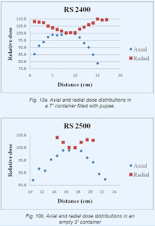
The 2-dimensional dose distribution within a container was measured by placing several long strips of film dosimeters within the container along the axis as well as along the diameter. The irradiated films were measured and the calibration was used to calculate dose values which were then expressed as a proportion of the central dose; the results are shown in figures 10a and 10b, for RS 2400 and RS 2500, respectively.

For RS 2400, this dose distribution was also measured by irradiating a large 14.5 x 17.2 cm Gafchromic sheet in the container filled with pupae. The irradiated dosimeter sheet was scanned in reflectance mode on an Epson 10000 XL flatbed scanner (model J181A) at 48 bits per pixel and 50 dpi. The calibration was used to calculate dose values which were then expressed as a proportion of the central dose (Fig. 11).

Dose uniformity for the 7” container for RS 2400 is better than ±15% over the container volume of 3.5 liters. While for 3” container for RS 2500, it is ±10% over the container volume of about 0.5 liters.


Conclusions
These new, self-shielded X-ray irradiators will fulfil the requirements of processing capacity, dose rate, dose uniformity and ease of use required for research and many small-scale applications, including sterile insect technique, small animal research, radiation resistant microorganism research, medical device terminal sterilization and viral inactivation. They, therefore, provide a practical alternative to self-shielded gamma ray irradiators with associated safety.

References
Kirk, R.E., Gorzen, D.F. 2008. X-ray tube with cylindrical anode. USA Patent number 7,346,147 B2.

National Academy of Sciences, 2008. Radiation source use and replacement: Abbreviated version. The National Academies Press, Washington. Available from: http://www.nap.edu/catalog/11976.html Accessed: Feb 5, 2010.

Rad Source Technologies Inc., 2009. RS 2400 - X-ray high volume irradiator. Available from: http://www.radsource.com/products/products.php?id=rs2400 Accessed: Nov 9, 2009.

Wagner, J.K., Dillon, J.A., Blythe, E.K., Ford, J.R., 2009. Dose characterization of the rad sourceTM 2400 X-ray irradiator for oyster pasteurization. Appl. Radiat. Isot. 67, 334-339.

Thursday, November 18, 2010

Practical X-ray Alternative To Self-shielded Gamma Irradiators

Kishor Mehta
Arbeiterstrandbad Strasse 72, Vienna A-1210, Austria
e-mail: mehta@aon.at

This is part one of the two part poster that was presented at the Health Physics Society annual meeting held in Salt Lake City, Utah, June 27th - July 1st, 2010. Part two will be coming soon. Select the link for the affiliated paper in Radiation Physics and Chemistry, Volume 80, Issue 1, January 2011, Pages 107-113.

Abstract
For high-energy photons (5-10 MeV), high powered X-ray irradiators have already been used for industrial applications, such as materials modification, food processing, and medical device sterilization. Now it seems possible to have smaller self-shielded irradiators with X radiation that can provide a very attractive alternative to self-shielded gamma ray irradiators. This has been made possible with the advent of the new technology, using 4π X-ray Emitters. The crucial characteristic of these emitters is a large distributed anode emitting photons in almost 4π geometry; this decreases the target cooling requirements, resulting in a higher power input and thus high dose rate. Of necessity, these irradiators have much smaller photon energies, maximum being 160 keV. However, the ultimate acceptance depends on the performance of these low-energy X-ray irradiators. The most important performance criteria for judging their success include dose rate, dose uniformity, throughput, reliability, safety and ease of operation. Another important requirement would be availability of dosimetry systems, reference as well as routine systems for dose measurements. Experimental data are presented here for two types of such self-shielded X-ray irradiators, both based on the 4π technology. These data include maximum dose rate achievable, dose uniformity ratio, the volumes that can be treated in one cycle and description of the dosimetry systems. The data indicate that these self-shielded X-ray irradiators are capable of replacing gamma irradiators for several applications, including sterile insect technique, small animal research, radiation resistant microorganism research, medical device terminal sterilization and viral inactivation.

Introduction
Self-shielded gamma ray irradiators (with cobalt-60 or caesium-137 as an isotopic source) have been extensively used for research and many small-scale applications, including sterile insect technique, small animal research, radiation resistant microorganism research, medical device terminal sterilization and viral inactivation. However it has now become increasingly difficult to purchase, transport or reload such irradiators because of the increased controls on radioisotopes due to the fear of terrorism. Fortunately, it now seems promising to have similar self-shielded irradiators with X radiation that can provide a very attractive alternative to self-shielded gamma ray irradiators. This has been made possible with the advent of the newly developed and patented technology (Kirk and Gorzen, 2008), using 4π X-ray emitters.

Since such irradiators do not depend on an isotopic source for the production of radiation, the danger of them being misused is eliminated, which seems to be the primary drive behind the move to replace gamma ray irradiators with X-ray irradiators (National Academy of Sciences, 2008 ). Before these devices can be used extensively and accepted by the research and scientific community, it is essential that its performance be demonstrated. Much experimental work has been done on two types of irradiators based on this technology. To add to some published reports (eg., Wagner et al., 2009), we present here a wealth of data that show suitability of these irradiators for the research purposes and some applications.

The data presented here are for two types of irradiators, namely RS 2400 Figure 1a and RS 2500 Figure 1b; both using 4π X-ray emitters.

RS 2400: The rotator system consists of five sample containers rotating around the X-ray emitter about 1.5 cm (variable) from the emitter surface in the fashion of a Ferris wheel, thus keeping their orientation fixed. The internal diameter and the length of the containers are 175 mm (variable) and 150 mm, resp. The wall of the container is made of ca. 1.4 mm carbon fibre reinforced resin, lined inside with 0.5-mm steel that acts as a spectrum-hardening filter. The volume of each container is about 3.5 litres giving a batch volume of about 17.5 litres.

RS 2500: It consists of one sample container surrounded by two X-ray emitters at a distance of about 1.5 cm (variable) from the surface. The container is placed on a table which can be rotated around its own axis. The internal diameter of the container is about 7 cm (variable) and the height can be varied according to the required dose uniformity in the sample.

X-ray irradiators
Each irradiator consists of two units: the irradiation chamber which contains the X-ray emitter(s), control system, power supply, and rotator with container(s) and the cooling unit which contains a water tank, pump and heat-exchanger for cooling the X-ray emitter(s). Figures 1a and 1b show the irradiation chambers, which are externally similar for RS 2400 and RS 2500 (Rad Source Technologies Inc., 2009).

The 4π X-ray emitter consists of an evacuated aluminium cylinder coated on the inside with gold (the X-ray converter) with the cathode arranged axially (Fig. 2). This cylinder is encased by a second cylinder to form a cooling water jacket. Photons are produced in 4π geometry; thus, they are emitted in all directions. This results in radiation field around the emitter consisting of photons contributed from all points along the anode. Typically the radiation field is more than twenty times that of a conventional point source, whilst the power density per unit area of target is much reduced.
Figures 3a and 3b show the arrangement of the sample container(s) and the X-ray emitter(s) for both types of irradiators. These geometries allow the sample to receive radiation from all directions in the rotation mode to improve dose uniformity. The characteristics are summarised in Table I.

Dosimetry systems
Reference dosimetry was carried out with a Farmer’s type 0.18 cm3 ionization chamber (supplied by RadCal Corporation, Monrovia, CA). It was calibrated in the photon energy range 50-1300 keV by the supplier with traceability to NIST (National Institute of Standards and Technology, MD, USA). Dose rate was measured at a reference location using this ionization chamber. Routine dosimetry was carried out using three types of radiochromic film dosimetry systems: two with Gafchromic® dosimeters and one with FWT dosimeters. The optical density (OD) was measured using a FWT Radiachromic® reader (FWT-92) for all the film dosimetry systems. For the calibration of these dosimetry systems, several dosimeters were irradiated at the reference location at each of several dose points in the desired dose range. Figures 4a and 4b show calibration curves for the Gafchromic DM 1260 dosimetry system and FWT dosimetry system.

Several experiments were carried out to study the effect of photon energy on the response of the Gafchromic film dosimeters. To change the photon spectrum at the centre of the container, the emitter was operated at 100 and 150 keV. The dose rate or (dose) was measured with the ionization chamber as well as with the Gafchromic dosimetry system (calibrated for 150 keV spectrum) for both these voltages. When exposed to the same field, the dose measured by the two sets of dosimeters differed by less than 3%, comparable with the uncertainty in the Gafchromic dosimetry system as well as the ionization chamber. Thus, it can be concluded that the response of the Gafchromic film dosimeters is energy independent in this photon energy range.

Wednesday, August 18, 2010

Revolutionary X-ray Tube Design

The 4 pi Emitter Series
The standard design for an X-ray tube has varied very little from the originals built by Roentgen in 1895. A beam of electrons was focused on a small target and cooling was applied to the back of the target. While this technology has worked well for applications such as medical imaging, which require minimizing radiation for safety purposes, it performs poorly in irradiation applications. The two primary reasons why X-rays have not been used for high radiation level applications are tube geometry and the inability to remove the heat generated during continuous operation.
In conventional X-ray tubes, the target emits a 4 pi field of photons at normal kVp levels (160 kVp). However, this field is limited by the tube geometry which only allows less than 18% of the photons generated to be used. The balance of the photons is absorbed into the shielding. In addition to the shielding that lowers the efficiency of the photon production, the X-ray spectrum itself must be filtered to achieve dose uniformity. To compensate for these two inefficiencies, the amount of current that generates the photons can be increased. Unfortunately, as the current is increased, so is the heat that is generated in the tube. The high current concentrated on a small target area (typically 1-3 kilowatts per square millimeter) is such that conventional X-ray tubes typically result in short tube life.
New patented technology has mitigated these shortcomings. Rad Source’s 4 pi emitter series uses entirely different geometry. A large cylindrical target reduces the heat resulting from the high current by vastly increasing the target area and thereby the power per unit area by a factor 4X10^5 (figure 1).
The same cylindrical target shape allows forward emitted electrons to go directly to the field and rearward projected photons to pass through the cylinder and contribute to the dose on the opposite side (figure 2). This results in a 4 pi field around the tube that utilizes 100% of the photon production as opposed to the less than 20% usage netted by conventional design.
In addition, the significantly reduced heat per unit area allows the use of a more efficient emission target. In conventional tubes a tungsten target is used because of its heat resistance. In this new design, the use of gold provides about a 10-15% increase in production efficiency and an even higher increase in average energy. This results in an X-ray emitter with about 20 times the dose per unit power of the conventional tube design.
The final improvement to the X-ray tube design is the ability to repair the tube. With any filament driven device, the filament will be consumed over time. The ability to replace the filament increases usable tube life many thousands of hours and reduces overall costs for irradiation. Currently the tubes are produced commercially up to 160 kVp. The International Atomic Energy Commission has evaluated equipment configured with Rad Source 4 pi emitter technology for Sterile Insect Technique (SIT) use and has designated it as an alternative to cobalt for SIT programs (IAEA, 2009). Other equipment incorporating this technology is being used by BSL-4 labs to achieve viral inactivation, providing dose rates up to 1 Megarad per hour. This is because the technology is scalable such that higher power, either as current or energy, can be increased with some design accommodation to meet customer’s needs. This technology is especially timely considering the nation’s current concerns regarding the potential use of radioactive materials for malevolent acts (U.S. Nuclear Regulatory Commission, 2006). To protect against such acts, many precautions have been taken and will be taken. Among these are tighter source restrictions, increased security requirements, and reduced disposal options, all of which has increased the cost of owning and using radioactive sources. Rad Source, anticipating these issues, has created alternatives to replace most self-shielded radioactive sources. To learn more about these alternatives please visit us at www.radsource.com or call us at 770-887-8669. About Rad Source Technologies Rad Source Technologies is currently the only company in the world supplying a comprehensive line of commercial X-ray radiation products designed to replace self-shielded gamma sources. The company’s current products are used for the irradiation of small animals, cells, sterile insect technique (SIT) applications, viral inactivation, and various other scientific applications.
###
IAEA. Joint FAO/IAEA Programme Nuclear Techniques in Food and Agriculture. Irradiation. Insect Pest Control Newsletter 72: 20; 2009. U.S. Nuclear Regulatory Commission. The Radiation Source Protection and Security Task Force Report. Report to the President and the U.S. Congress Under Public Law 109-58, The Energy Policy Act of 2005 vii; 2006.

Thursday, July 15, 2010

Gamma vs X-ray Comparison

What is Ionizing Radiation?
Ionizing radiation consists of subatomic particles or electromagnetic waves that are energetic enough to detach electrons from atoms or molecules, ionizing them. Two of the most common forms of ionizing radiation are gamma rays and X-rays. Both forms of ionizing radiation are almost identical with exception to their source of origination. Gamma rays originate from the nucleus while X-rays originate in the electron fields surrounding the nucleus or are machine produced.

How is Ionizing Radiation Generated?
Ionizing radiation comes from radioactive sources such as cobalt-60 and cesium-137 and non-radioactive sources such as X-ray tubes. Radioactive sources are unstable materials that generate gamma rays as they decay. X-rays are generated in a vacuum tube where high voltage is used to accelerate electrons to a high velocity, that then collide with a metal target, an anode creating X-rays.

How is X-ray and Gamma Ionizing Radiation Different?
There are three primary differences between X-ray and gamma ionizing radiation; frequency, wavelength, and photon energy. While the first two are used as identifiers to differentiate the various wavelengths, the third, photon energy describes the energy or speed at which the rays are traveling. Described in units of electronvolts, cesium-137 is 662 keV, cobalt-60 is 2.5 MeV, and Rad Source X-ray is 160 kVp. This energy equates to penetration power; the higher the energy the greater the penetration power. For both gamma and X-ray, energies are emissions in free space. In actual use, where they are confined in a lead chamber, the energies of both are affected by scattering and fluorescence until actual energy spectrums are difficult to define. The greater the energy, the more shielding is required for safe operation.

What is Ionizing Radiation Used for?
Because of the penetrating properties of ionizing radiation and their ability to inactivate microorganisms, ionizing radiation is used for a number of different purposes. Including, virus inactivation for research, as well as to sterilize or reduce the microbial load of many different types of products such as medical devices, packaging, cosmetics, foods, and agricultural products. It is also used to alter the properties of many different polymers through recombination, cross-linkage, and cross scission.

Why use X-ray versus Gamma produced Ionizing Radiation?
Radioactive sources are very dangerous requiring specialized shipping containers and services with heavy shielding and high levels of security. The radioactive sources once delivered, require specialized rooms and personnel must have background checks, and radiation badges to operate the radioactive unit. The unstable material is constantly decaying and cannot be turned off. Cobalt-60’s half-life is 5.27 years while cesium-137’s half-life is 30.17 years. Once the radiation source drops below a useable level, the radioactive source generally cannot be reloaded and must be disposed of following specific, costly protocols that involve the above mentioned shipping containers and the long-term storage of the radioactive source that continues to degrade for tens to hundreds of years.

X-ray ionizing radiation is produced by a X-ray tube, therefore it can be turned off when it is not in use. At 160 kVp, X-rays have more than enough penetration power to achieve the desired results and yet requires much less shielding. The X-ray unit does not require any special licensing or special room accommodations*. Operators of the X-ray unit do not require background checks prior to operation nor do they require the use of radiation badges (21CFR1020.40 compliant). When the X-ray unit reaches the end of it’s lifecycle, it does not require the expensive disposal costs associated with the transportation and storage of radioactive sources.

*Based on using a Rad Source Technologies unit and current
operating knowledge of global regulations. This information is subject
to change, please contact your Rad Source representative for specific
and up-to-date information.

This information is provided by Rad Source Technologies. The full document can be found at Gamma vs X-ray Comparison. If you have any questions, please contact Rad Source at 770.887.8669 or contact us.

Friday, July 9, 2010

Food Irradiation in the News

Below are links to news articles discussing food irradiation research performed by Dr. Barakat S.M. Mahmoud of Mississippi State University using the Rad Source RS 2400 which features the revolutionary 4 pi X-ray emitter technology.
Contact information for Dr. Barakat S.M. Mahmoud at Mississippi State University
Enhanced by Zemanta

Thursday, July 8, 2010

Rad Source at the 55th HPS Annual and 22nd Biennial Campus Radiation Safety Officers Meeting

SlcoldImage via Wikipedia

Rad Source Technologies hosted an exhibitor booth at the 55th HPS Annual and 22nd Biennial Campus Radiation Safety Officers Meeting held this year in Salt Lake City, UT. The exhibitor portion of the meeting was held at the Salt Palace Convention Center, June 28th - 30th, and again provided Rad Source the opportunity to promote our innovative 4 pi emitter X-ray tubes and their high dose irradiation applications. Again, thanks to all who visited our booth. If you are interested getting more information about the 4 pi technology, the applications, or anything else that has to do with self-shielded gamma irradiator replacement please submit your inquiry to contact us.

About Rad Source Technologies
Rad Source Technologies is currently the only company in the world supplying a comprehensive line of commercial X-ray radiation products designed to replace self-shielded gamma sources. The company’s current products are used for the myeloablation of small animals, cells, sterile insect technique (SIT) applications, virus inactivation, food irradiation research, seed mutation, and various other scientific applications.
Enhanced by Zemanta